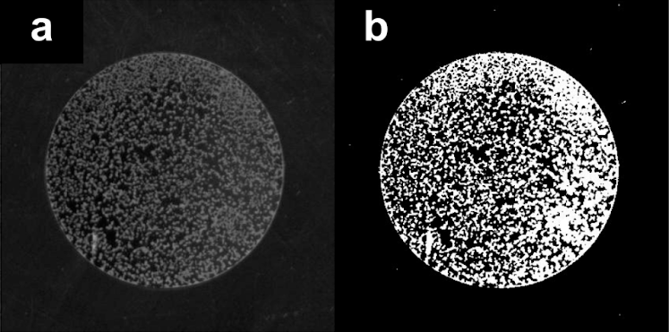
PA quantification: (a) Preprocessed image of one well on a 96-well plate containing an S. cerevisiae strain after growth in liquid SLD, then washed; (b) Processed by computational pipeline.
PA was phenotyped using synthetic low dextrose (SLD) media (0.67% yeast nitrogen base without amino acids and with ammonium sulfate and 0.1% dextrose). After 2 days of 30C incubation, plates were emptied, rinsed 3 times with DI water, and the remaining adherent cells were scanned using an Epson Expression 11000 XL scanner.
A Python pipeline was used to process the images of the bottom of PA wells after washing away culture and non-adherent cells. A threshold was applied to the image to divide the image binarily: (1) yeast cells, (2) background. PA was quantified using the number of adhering yeast cells (white pixels in b) divided by the total number of pixels (black and white in b) at the bottom of the well, represented by the circles in the image.